
Your body isn't broken.
Pain is an output. Not a damage report.
You've had the scans, the referrals, and the ice packs. Nothing is wrong — but it still hurts. Modern pain science says the answer isn't in your tissues. It's in your nervous system.
You’ve done the right things. You went to the doctor. Got the MRI. Saw the specialist. Maybe two. You’ve tried the stretches, the ice, the ibuprofen, the foam roller, the cortisone shot. Some of it helped for a while. Most of it didn’t.
And somewhere in that process, somebody looked at you and said one of two things: “there’s nothing wrong,” or “we found something on the scan.” Neither one made the pain make sense. Because if nothing is wrong, why does it hurt? And if the scan explains it, why didn’t the treatment fix it?
Here’s what I want you to consider: your body probably isn’t broken. The pain is real. It’s not in your head. But it may not be coming from where you think.
The image that didn’t tell the whole story.
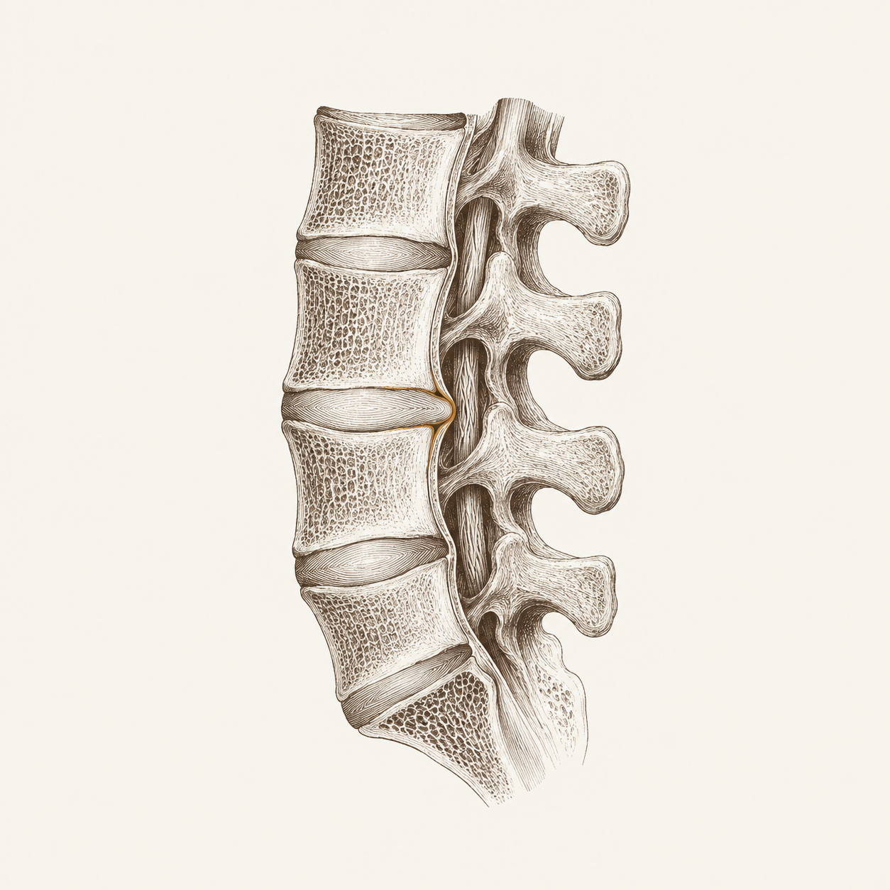
In 2015, a team of researchers did something simple. They pulled together every study they could find on spinal MRI findings in people with no symptoms at all — no back pain, no complaints, nothing. Just routine imaging. They looked at 33 studies, covering more than 3,000 people.1
What they found should have changed the conversation. Disc degeneration showed up in 37% of 20-year-olds with no pain. By age 80, 96% had it. Disc bulges appeared in 30% of 20-year-olds. Disc protrusions in 29%. These aren’t injuries. They’re structural variation — as common and unremarkable as grey hair, and just as weakly correlated with how you actually feel.
This doesn’t mean imaging is useless. If you’ve had a trauma, a fall, new neurological symptoms — you need the scan. But for the person who’s been hurting for months, got an MRI, and was told their disc is the reason: the evidence suggests that disc was probably there before the pain started, and it’ll be there after the pain stops.
The image isn’t the story. The nervous system is.
Pain is not a damage report.
Here’s where it gets interesting. In 2020, the International Association for the Study of Pain — the organization that literally defines the word — updated their definition. Pain is now described as “an unpleasant sensory and emotional experience associated with, or resembling that associated with, actual or potential tissue damage.”2
Read that again. Or potential. The definition itself says pain does not require damage. Pain can occur when the brain predicts threat, even when no tissue has been harmed.
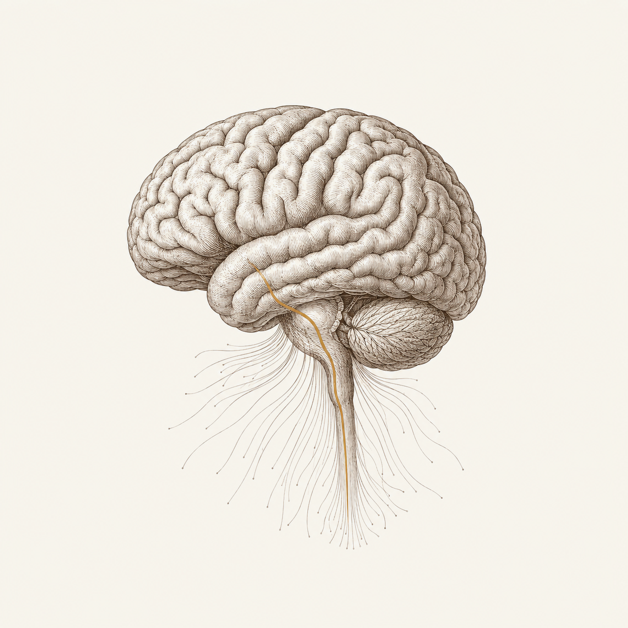
This isn’t fringe. This is the consensus scientific definition, adopted by the organization that sets the standard. Pain researchers have been saying some version of this for decades: pain is an output of the brain, not an input from the body.3 The brain takes in information from the tissues, yes — but also from your history, your beliefs, your stress level, the context you’re in, your sleep, your sense of safety. It weighs all of it. And it decides whether to produce pain.
That decision can be wrong. The brain can produce pain when the tissues are fine. And it can suppress pain when the tissues are damaged — soldiers in combat, athletes mid-game. The relationship between tissue state and pain experience is real but loose, not the tight one-to-one you’d expect.
The alarm that stays on.
So the brain can produce pain without damage. But why would it keep producing it?
Central sensitization is the mechanism that explains most persistent pain that outlasts the original injury.4 Here’s what happens: an injury occurs. Nociceptors — the nerve endings that detect potential threat — send signals to the spinal cord and brain. The system responds appropriately. But if the signals persist long enough or intensely enough, the spinal cord and brain undergo a change. The gain gets turned up. Neurons in the dorsal horn become hyperexcitable. Signals that used to be subthreshold now cross it. Touch that was fine before now registers as painful. Movement that was tolerable becomes sharp.
The alarm volume got turned up. And then the injury healed. But the volume stayed.
This is real neural plasticity — the same kind of plasticity that lets you learn a language or develop a skill, running in the wrong direction. The system got better at producing pain.5 And it doesn’t automatically reset when the tissue does. There’s no timer. No automatic return to baseline. The sensitized state can persist for months or years after the original event has fully healed.
If you’ve ever had pain that started with something specific — a strain, a tweak, a bad day at the gym — and then just… didn’t leave, long after the tissue should have healed: this is likely what happened. The tissues recovered. The alarm didn’t.

The state that keeps the alarm sensitive.
Here’s where the work I do connects to the pain science.
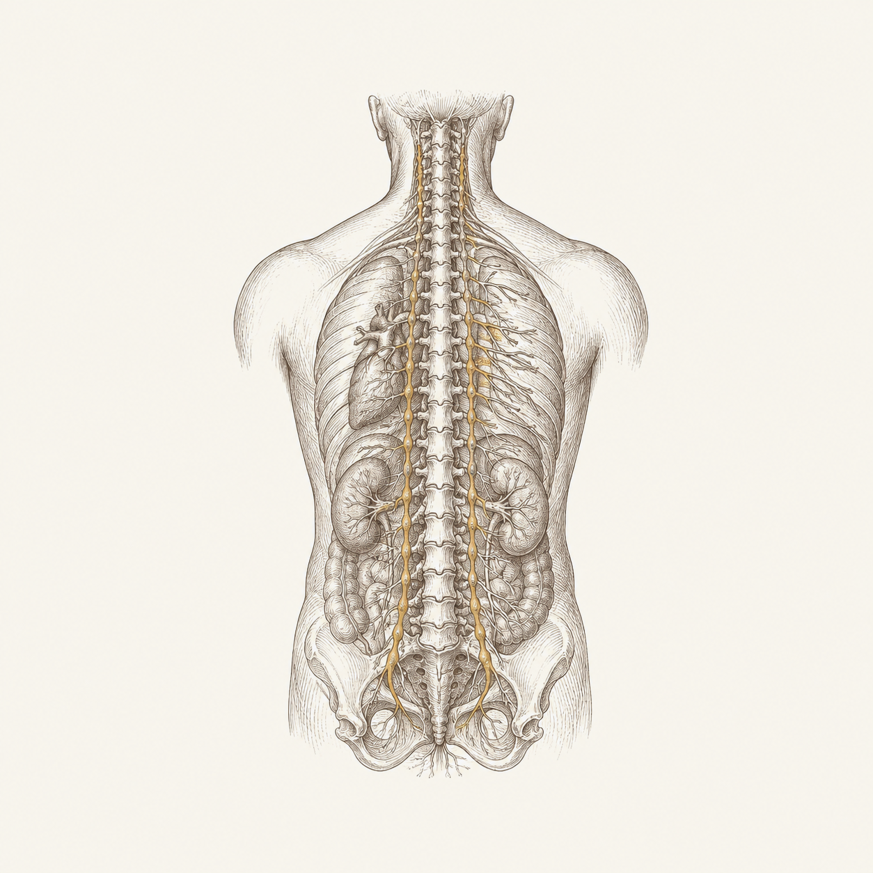
The autonomic nervous system — the system that runs your stress response, your digestion, your heart rate, your breathing — doesn’t just sit next to the pain system. It modulates it. Sympathetic dominance (the “on” state, the alert state, the state that keeps you braced and ready) lowers the threshold for pain. It makes the alarm easier to trip.†
This is the same state I’ve written about in the context of sleep (#17), recovery (#15), and training (#14). The pattern is consistent: when the nervous system is stuck in a sympathetically dominant position, everything downstream is affected — sleep quality, recovery capacity, training response, and pain sensitivity.†
The research on slow breathing and autonomic tone is relevant here. Exhale-focused breathing shifts the autonomic balance toward the parasympathetic side.6 It lengthens vagal output. It lowers the baseline. And in clinical practice, when that shift happens, pain often changes — sometimes immediately, sometimes over days.†
Not because breathing cures the disc, or the tendon, or whatever the scan showed. But because the state that was amplifying the signal quiets down, and the actual tissue state — which may have been fine all along — gets a chance to report accurately.
The honest version.
The MRI evidence is the strongest piece here. The asymptomatic-imaging literature is large, replicated, and largely uncontroversial among researchers — the disconnect between structural findings and symptoms is well-established.1 If you take one thing from this piece, let it be that.
Central sensitization is well-documented as a mechanism.45 The evidence that it explains a significant portion of persistent non-specific pain is strong and growing. The clinical challenge — determining which patient’s pain is centrally driven vs. peripherally driven — is harder, and honest clinicians will tell you there’s no clean biomarker for it yet.
The autonomic connection — the idea that sympathetic dominance lowers pain thresholds and that respiratory intervention can modulate the state — is built on established mechanism (slow breathing shifts autonomic tone6) connected to a clinical observation (pain changes when the state changes†). The connection is coherent and I see it consistently. It is not a controlled finding from a pain trial. I’m applying respiratory physiology to a pain context, which is my clinical reasoning, not a replicated result.
The positional pattern interpretation — that specific postural and respiratory patterns maintain the autonomic state that amplifies pain — is the most framework-dependent claim.† It’s how I read it. It guides the assessment and the intervention. But it hasn’t been tested in a study designed to isolate it from the respiratory and autonomic effects, and I’d rather say that than pretend it has.
What I can tell you: people walk in with pain that hasn’t responded to anything structural, and when we address the nervous system state through positional respiratory work, the pain changes. Not always. Not everyone. But consistently enough that I stopped thinking of it as coincidence a long time ago.
If you’ve had pain that doesn’t match a scan, doesn’t respond to the usual fixes, and nobody’s been able to explain — an assessment might show you something that imaging can’t. Not what’s broken. What state your system is in, and why it’s staying there.
Book an assessment at aerboston.com
The fine print
-
Brinjikji W, Luetmer PH, Comstock B, et al. Systematic literature review of imaging features of spinal degeneration in asymptomatic populations. AJNR Am J Neuroradiol. 2015;36(4):811–816. doi:10.3174/ajnr.A4173. Limitation: a systematic review of cross-sectional imaging studies. It establishes prevalence of structural findings in asymptomatic people, not whether those findings will later become symptomatic. The included studies vary in imaging protocols and population demographics. The finding that structural degeneration is common without pain is robust; the implication that a given patient’s scan finding is “incidental” in any individual case requires clinical judgment.
-
Raja SN, Carr DB, Cohen M, et al. The revised International Association for the Study of Pain definition of pain: concepts, challenges, and compromises. Pain. 2020;161(9):1976–1982. doi:10.1097/j.pain.0000000000001939. Limitation: a consensus definition, not an original research finding. It reflects the current expert understanding but is a product of committee negotiation. The inclusion of “or potential tissue damage” and the accompanying note that pain can occur without tissue damage codify a position held in pain science for decades, now formalized.
-
Moseley GL, Butler DS. Fifteen years of Explaining Pain: the past, present, and future. J Pain. 2015;16(9):807–813. doi:10.1016/j.jpain.2015.05.005. Limitation: a perspective/review piece, not an original trial. It summarizes the Explain Pain approach and its evidence base, including trials of pain neuroscience education that show modest improvements in pain and disability. The therapeutic implication (that understanding pain changes the pain experience) is supported but effect sizes are moderate and variable across populations.
-
Woolf CJ. Central sensitization: implications for the diagnosis and treatment of pain. Pain. 2011;152(3 Suppl):S2–S15. doi:10.1016/j.pain.2010.09.030. Limitation: a review and conceptual framework paper. Central sensitization as a neural mechanism is well-established in animal and human studies. The clinical challenge — reliably distinguishing centrally maintained pain from peripheral nociceptive pain in a given patient — remains an active research problem. No validated clinical biomarker exists for central sensitization specifically.
-
Latremoliere A, Woolf CJ. Central sensitization: a generator of pain hypersensitivity by central neural plasticity. J Pain. 2009;10(9):895–926. doi:10.1016/j.jpain.2009.06.012. Limitation: a comprehensive review of the neural mechanisms. The molecular and cellular evidence for central sensitization is strong. Translation from mechanism to individual clinical diagnosis is less established — the review itself focuses on the biological basis, not on clinical application.
-
Laborde S, Allen MS, Borges U, et al. Effects of voluntary slow breathing on heart rate and heart rate variability: a systematic review and a meta-analysis. Neurosci Biobehav Rev. 2022;138:104711. Limitation: the vagal effects are clearest during and shortly after the practice; evidence that slow breathing durably shifts resting autonomic tone is thinner and more mixed. Applying slow breathing to a pain modulation context specifically is our reasoning based on the autonomic mechanism, not a result from a pain-focused trial.
The Letter
The writing here is the why. The letter is where the what to do lives — the protocol, the drill, the case behind each piece.
If any of this changed how you think about your own body, an assessment is where that conversation starts.