
You can only breathe on one side.
The asymmetry that feeds itself 20,000 times a day.
You checked your shoes. You noticed your sleep side. Now put your hands on your ribs and breathe. One side moves more than the other. That's not a habit — it's the same structural asymmetry, playing out in every breath you take.
Put your hands on your ribs. Both sides, fingers spread. Breathe in.
Notice which side moves more.
If you’ve been reading the last two of these, you already know which side it is. It’s the same side that carries your weight. The same side whose shoe wears out faster. The same side you roll toward in bed.
That’s not a coincidence. It’s the same pattern — the same structural asymmetry we’ve been tracing since No. 21 — but now it’s in your breathing. And that matters more than the shoe or the sleep side, because this one reinforces itself with every single breath you take.
About 20,000 times a day.
The diaphragm has a side, too.
We covered this in broad strokes in No. 21. Your body’s internal architecture is not symmetrical — three-pound liver on the right, heart on the left, three lung lobes right, two on the left. The diaphragm sits on top of all of that, and it inherits every bit of the asymmetry.1
Here’s the detail that matters for breathing.
The right side of your diaphragm is larger. It has more muscle fibers. It sits higher in the chest at rest, because the liver — dense, firm, incompressible — pushes it up from below like a platform. The left side sits lower, shares space with the heart, and has nothing that solid underneath it. Below the left hemidiaphragm: the stomach, the spleen. Soft, compressible organs that shift with position and with the meal you ate two hours ago.1
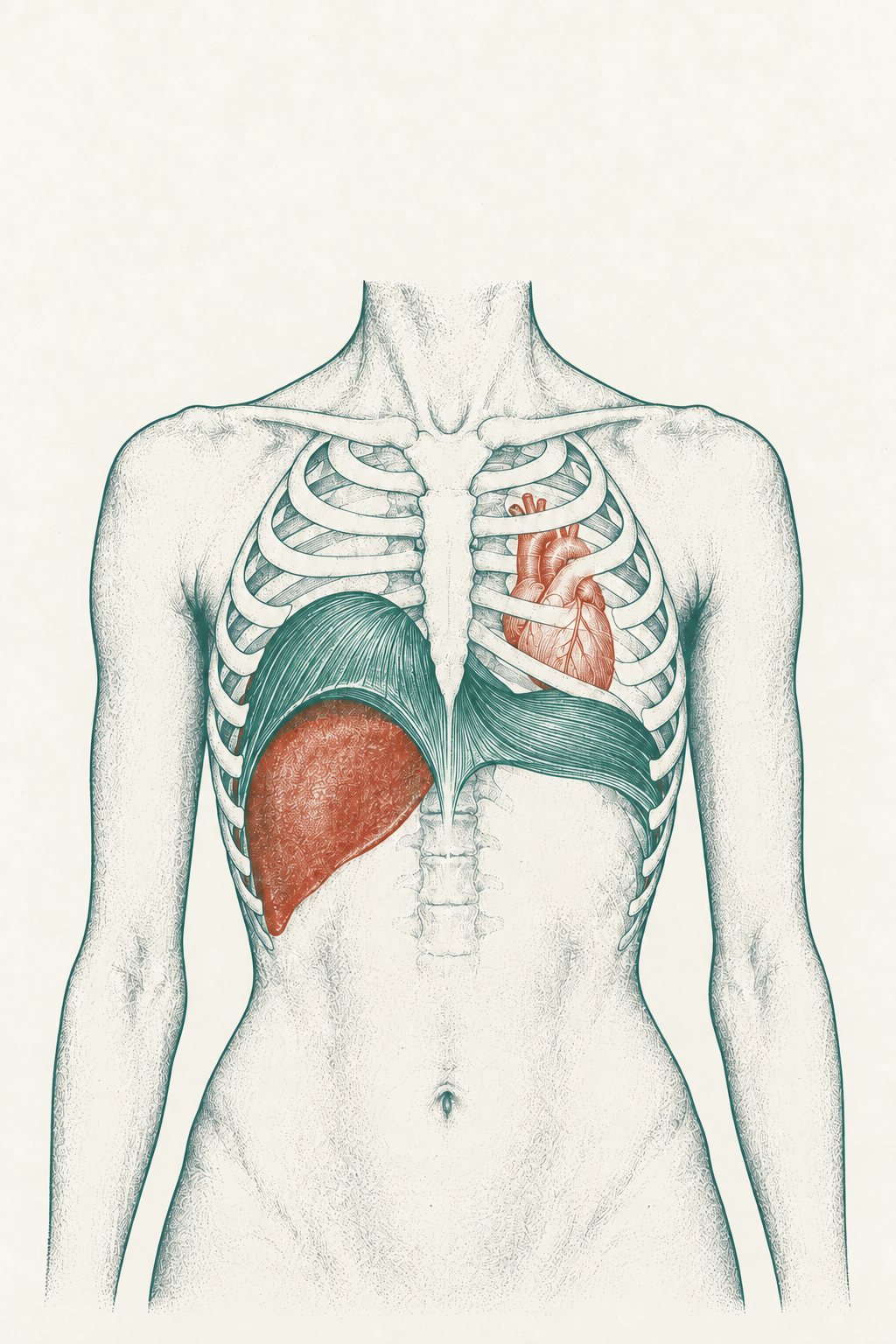
This isn’t subtle. On a chest X-ray, the right hemidiaphragm normally sits visibly higher than the left.2 That height difference means more starting length for the muscle fibers on the right — and a longer muscle at rest contracts more forcefully. The right side of your diaphragm has better mechanical advantage before you take a single breath.
What the left side loses.
Here’s where it gets interesting — and where the clinical framework starts.†
Your diaphragm doesn’t just pull air in. It stabilizes your trunk. The part of the diaphragm that matters for both jobs is the zone where the muscle fibers run vertically along the inside of your lower ribs — apposed to the ribcage, pressed against it, creating intra-abdominal pressure that holds your trunk stable. In PRI terminology, this is the Zone of Apposition.†
When the ZOA is intact — when the diaphragm maintains its dome shape on both sides — the muscle can do both jobs. It descends on inhale, pulling air into the lungs. It pushes against the ribcage wall, stabilizing the trunk. Both sides contribute. You breathe into both sides of your chest. Your trunk is stable from both sides.†
But the left side is vulnerable. Without the liver underneath it, the left hemidiaphragm tends to lose its dome. It flattens. The muscle fibers that should run vertically along the ribs become more horizontal — more like a flat sheet than an arched dome. When that happens, the Zone of Apposition collapses on the left.†
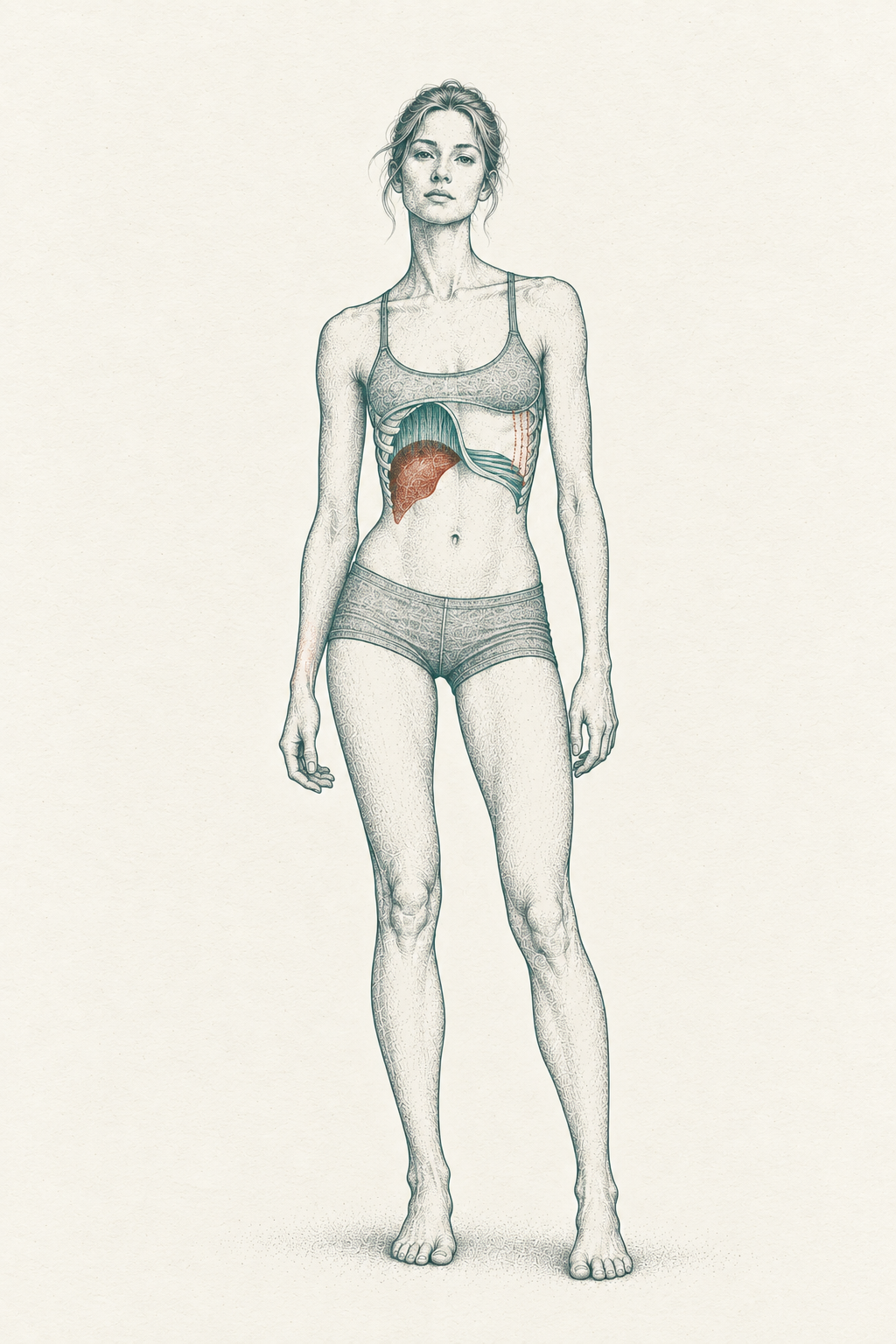
A flat diaphragm can’t descend much further — it’s already descended. So the left side of the diaphragm can’t pull air in as effectively. And it can’t stabilize the left side of the trunk as effectively. One muscle, two jobs lost.†
What this feels like.
The right side of your chest expands more easily. More air goes right. The left side feels tighter, harder to fill, restricted. If you’ve ever felt like you can’t get a full breath, or that one side of your chest is stuck — that’s the architecture playing out in real time.
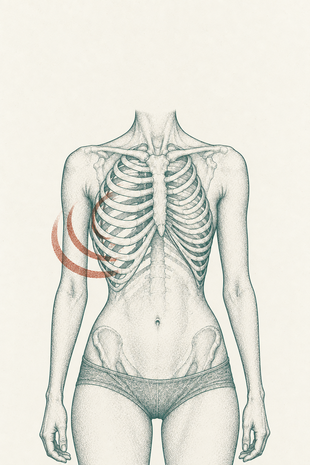
The research supports this, independent of any clinical framework. Studies measuring regional lung ventilation show that in most body positions, ventilation distributes asymmetrically — the dependent lung (the side closest to the ground) receives more perfusion, and positional changes shift ventilation from one side to the other.3 Your breathing is position-dependent, and if your resting position favors one side, your breathing follows.
If you train hard and track your numbers, you might notice this from the other direction. Heavy unilateral work — single-leg deadlifts, lunges, loaded carries — biases your system further into the pattern. The side that’s already working harder works harder still. And the breathing pattern that feeds the postural pattern gets another 20,000 repetitions to reinforce it.†
Your HRV might dip the day after heavy one-sided loading — not because you overtrained, but because the autonomic system reflects the asymmetry in respiratory mechanics. That’s a hypothesis, not a proven mechanism.† But the athletes and high-performers who track everything often notice the pattern before they understand it.
The feedback loop.
This is what makes the breathing piece different from the shoe wear or the sleep side. Those are outputs of the pattern. Breathing is both an output and an input.†
The left diaphragm loses its dome → the left chest doesn’t expand → less air goes left → the left side of the trunk loses stability → the system leans further into the right side → the left diaphragm flattens more → the left chest expands even less.†
Twenty thousand breaths a day, feeding the spiral.
Your worn-out shoe tells you where the pattern went. Your breathing is where the pattern lives. It’s the engine.†
What you can feel right now.
Lie on your left side. Knees stacked, bent to about 90 degrees. Put your right hand on the right side of your ribcage — the side now facing the ceiling.
Breathe in through your nose. Feel the ribs push into your hand. Exhale fully through your mouth. Do five breaths.
Now flip. Lie on your right side. Left hand on the left ribs — the side now facing the ceiling.
Same thing. Five breaths. Feel what happens.
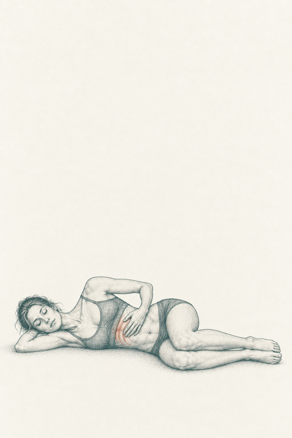
For most people, one side moves more. The ribs push into the hand more easily. The breath fills more completely. The other side feels stuck — like the ribs don’t want to move, like the air can’t get there, like you’re breathing through a wall.
That’s the asymmetry. Not in your shoes. Not in your mattress. In the architecture of the breath itself.
The pattern feeds itself through every breath. The worn shoe records it. The sleep side accommodates it. But the diaphragm drives it — 20,000 cycles a day, reinforcing the same side, deepening the same spiral.
That’s what an assessment finds. Not just which side is tight. Where the breathing pattern sits, how the diaphragm is organized, and what it takes to get the other side back online.†
Next week: the same side that owns your breathing shows up in your jaw, your neck, and your eyes. No. 24 traces the chain.
Book an assessment at aerboston.com
The fine print
-
Standring S, ed. Gray’s Anatomy: The Anatomical Basis of Clinical Practice. 42nd ed. Elsevier; 2021. Chapters on the thorax and diaphragm. The structural asymmetry of the diaphragm — right hemidiaphragm larger, with more muscle fibers, sitting higher due to the liver; left hemidiaphragm smaller and sharing the thoracic cavity with the heart and softer abdominal viscera — is standard human anatomy. The same source verified in No. 21.
-
Nason LK, Walker CM, McNeeley MF, Burivong W, Fligner CL, Godwin JD. Imaging of the diaphragm: anatomy and function. RadioGraphics. 2012;32(2):E51-E70. doi:10.1148/rg.322115127. PMID: 22411950. States that “the right hemidiaphragm is normally slightly higher than the left hemidiaphragm.” A comprehensive review of diaphragm anatomy and function using imaging modalities. Limitation: a narrative review, not an original measurement study; characterizes the asymmetry as “slight” without quantifying the difference in intercostal spaces.
-
Petersson J, Glenny RW. Gas exchange and ventilation-perfusion relationships in the lung. Eur Respir J. 2014;44(4):1023-1041. doi:10.1183/09031936.00037014. PMID: 25063240. A comprehensive review of ventilation-perfusion relationships in the lung, including how V/Q mismatch arises in various conditions. The well-established finding that ventilation and perfusion distribute non-uniformly in the lung is a foundation of respiratory physiology; the same authors have demonstrated in experimental work (Petersson et al., Respir Physiol Neurobiol, 2007; PMID: 17169620) that posture changes redistribute both ventilation and perfusion toward the dependent lung. Limitation: a review article synthesizing prior work; the specific claim about resting postural bias driving habitual ventilation asymmetry is my inference, not a conclusion of this paper or the broader literature.
The Letter
The writing here is the why. The letter is where the what to do lives — the protocol, the drill, the case behind each piece.
If any of this changed how you think about your own body, an assessment is where that conversation starts.